
Eriophyid mites are distributed ubiquitously in Bulgaria and damage grapevine, fruit trees, small fruits, vegetable, field and other crops. Many species feed and develop on weeds, some exhibit high specificity and are used for biological control (Stoeva A., V. Kharizanova, 2009). Eriophyid mites are invisible without magnification. Many species lead a concealed way of life, and their damage is often attributed to other causes. This makes them unknown or little known to specialists and producers. The first scientific data on them date back to the middle of the 19th century, and during its last quarter significant scientific research has been conducted on species composition, morphology, systematics and their harmful activity. So far about 3600 species are known in the world eriophyid mite fauna, and for the Bulgarian fauna – 129 (Stoeva A., V. Kharizanova, 2009). In Bulgaria, the main studies on this group of mites have been conducted by Prof. Dr. P. Nachev (Raykov E., P. Nachev, 1957; Nachev P., 1963, 1967, 1976, 1979; Raykov E., P. Nachev, 1971; Balevski A., P. Nachev, Sp. Simova, 1980; Nachev, 2008 and others), and information on the harmful activity and biology of individual species has been reported by Kharizanov A., L. Georgiev (1972), Kharizanov A., Iv. Molenin, P. Abrasheva (1980), Kharizanov et al. (1994), Kharizanov (2011) and others.
Eriophyid mites belong to cl. Arachnida, subcl. Acarina, ord. Acariformes, subord. Prostigmata, superfamily Eriophyoidea and families Eriophydae, Nalepellidae and Rhyncaphytoptidae.
Fam. Eriophydae – the chelicerae are short (15–40 µ), the dorsal shield lacks anterior dorsal setae. The first pair of legs has no tibial spine, the genital coverflap is striated.
Fam. Nalepellidae – the chelicerae are very short, the dorsal shield bears one or two setae, directed mainly forwards. On the tibia of the first pair of legs there is a spine located laterally, and the genital coverflap is smooth.
Fam. Rhyncaphytoptidae – the chelicerae are long and in some species reach 70 µ. The dorsal shield bears setae directed forwards or inwards, and the genital coverflap is striated. The family includes the largest and free-living species.
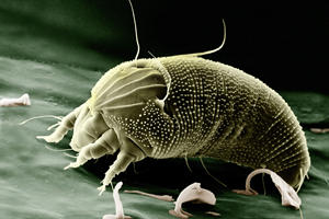
Brief morphological characteristics. The body of eriophyid mites is elongated, vermiform, cylindrical, spindle-shaped, pointed and varying in colour – white, pale yellowish, yellow-brown, rusty-brown, grey-brownish or otherwise coloured. Its length in the individual species varies from 100 to 304 µ; width – from 40 to 80, and thickness – from 50 to 88 µ. It consists of 3 tagmata – gnathosoma (rostrum, proboscis), propodosoma and hysterosoma. The legs are two pairs and are articulated to the first and second body segments. The gnathosoma consists of the chelicerae and pedipalps. The latter are fused with the basal parts of the first pair of legs and form the so-called rostrum, composed of basal, intermediate and terminal segment. In its anterior part there is a canal in which the chelicerae are placed, and in the basal segment – fused with the segments of the first pair of legs – are located the pharynx and the so-called pharyngeal pump. The chelicerae are needle-shaped, straight or slightly curved, pointed anteriorly and broadened at the posterior end, which is rounded and by rhythmic rotations moves the chelicerae forwards and backwards. Behind the chelicerae is the so-called oral stylet (similar to the chelicerae), which conducts saliva into the cells of plant tissues and takes up plant sap. Feeding is carried out by contraction of the pedipalps, whose segments are connected telescopically. The propodosoma is short and occupies a small part of the body. The dorsal side is occupied by the so-called dorsal shield, and the ventral side – by the basal segments of the legs. The dorsal shield bears various structures used as specific morphological characters. The legs are composed of trochanter, femur, genu, tibia and tarsus, which also bear specific morphological characters. The hysterosoma occupies the largest part of the body and is most often cylindrical in shape. It is transversely striated, composed of separate rings, whose number and shape are important morphological characters. In species living in galls, buds and other concealed sites, the annular formations are identical on the dorsal and ventral sides, but broader than the ventral ones and with specific outgrowths. The last 4–6 segments are called telosoma and are identical in the two groups of mites. The terminal ones resemble a “sucker” and secrete a volatile substance facilitating mite movement. The hysterosoma is also covered with many setae which characterize the chaetotaxy of the species. At the anterior end of the ventral side, behind the basal segments of the second pair of legs, the reproductive system is located. The genital opening is transversely positioned and covered by a plate – the genital coverflap. It has thickened edges forming a chitinized frame around the genital opening and an internal skeleton, specific for the individual species.
Damage symptoms. Eriophyid mites suck sap from various parts of plants, secrete saliva (enzymes) and induce anatomical-morphological, physiological and biochemical changes in the tissues, which manifest as speckling, deformations, overgrowth, gall formation and other changes.
- Leaf chlorosis – the damaged leaves have a silvery sheen, since the sap from the epidermal cells has been sucked out, their interior is filled with air, and the cell walls have retained their integrity.
- Rusting (browning) of leaves – the cell sap of the epidermal cells is sucked out, but the cell walls shrink and become necrotic – they turn brown. Such cells have a delicate cell wall and high water content and are typical of tender and rapidly growing plant parts (leaves and fruits) – most often in vegetable crops, grapevine and others. Deformation – expressed as curling or crinkling of the leaf blade and bending of the midrib (as a result of abnormal growth) and as changes in the floral organs.
- Erineum formation is also a deformation of the leaves affecting the epidermal cells. During sap sucking the mites secrete enzymes that cause strong elongation and thinning of epidermal cells, turning them into cotton-like hairs. They are mainly on the underside of the leaf blade, initially white, but later turn brown and black. On the upper side the tissues are raised, and on the underside – depressed. The mites live and feed between the hairs.
- Gall formation – galls are closed formations resulting from tissue overgrowth with various shape and size – conical, on the upper or lower leaf surface, or rounded on the bark at the base of young shoots. The inner walls of the galls are succulent and often lined with erineum.
- Bud overgrowth – damaged buds overgrow, become deformed and change their shape and colour. Eriophyid mites are also vectors of viruses causing diseases in small fruits, cereals and other crops. Life cycle. The life cycle of eriophyid mites passes through the stages egg, protonymph, deutonymph and adult mite. The larva develops in the egg, moults and the pronymph hatches. Mites living in the open develop two forms of females – protogynes (summer form) and deutogynes (winter form). Protogynes develop from the end of spring to the beginning of autumn, and deutogynes – from the beginning of autumn to the end of spring. Protogynes live about 30–35 days, after which protogynes of the next generation appear. They are followed by males, which provide spermatophores for fertilization of the females. Deutogynes differ from protogynes by a coarser body integument, weakly expressed tubercles, structural formations on the dorsal shield and a darker colouration. They are significantly more resistant to unfavourable abiotic factors than protogynes. According to Shevchenko et al. (1968), males also appear among the deutogynes. Protogynes develop and cause damage on shoot tips and other young and growing plant parts, and after these parts age, deutogynes appear on them, cause damage and develop.
Eriophyid mites develop from 1 to 10–15 generations per year and overwinter as females in buds, galls, crevices and cracks, in galls on the bark at the base of twigs and in other protected sites. There are species that develop without diapause, which complete one generation in 5–7 days.
Eriophyid mites are controlled by various species of predatory insects, mites and spiders, by heavy rains, wind and low temperatures, and are sensitive to systemic organophosphorus insecticides, neonicotinoids and acaricides.

